It is a heavy Saturday at an orthopaedic hospital in Delhi NCR. The X-ray queue is long, the on-duty radiologist has read more than a hundred films, and a hairline scaphoid fracture on a young patient's wrist slips through unread. The patient goes home, the pain is dismissed as a sprain, and weeks later the missed injury becomes a complication, a complaint, and a question no hospital wants to answer.
This is the quiet cost of diagnostic error in orthopaedics, and it is where AI medical imaging solutions are starting to help. By acting as a consistent second set of eyes on every scan, they help hospitals catch what fatigue and volume cause humans to miss. The sections below cover how this technology works for orthopaedic care, how to reduce diagnostic errors in orthopaedics with AI, and what it takes to deploy it in line with Indian regulation.
Why Diagnostic Errors in Orthopaedic Imaging Are So Costly
Missed fractures are one of the most persistent failure points in orthopaedic and trauma care. Subtle, hairline, and occult fractures are easy to overlook on a plain radiograph, especially in busy emergency settings where reader fatigue and time pressure are constant. The challenge grows with India's shortage of trained radiologists, which leaves fewer specialists reading more scans than ever.
The consequences are not only clinical. A missed or delayed diagnosis can mean the wrong treatment, a prolonged recovery, an avoidable second procedure, and rising medico-legal exposure. It also erodes a speciality hospital's reputation for accuracy.
For decision-makers weighing AI in orthopaedic diagnostic imaging, the problem usually comes down to a few recurring pressure points:
- Subtle and occult fractures missed on first read
- A growing imaging backlog and slow turnaround times
- Reader fatigue during high-volume shifts
- Medico-legal risk from delayed or incorrect diagnosis
How AI Medical Imaging Solutions Work for Orthopaedic Care
At their core, these systems use computer vision for diagnostic imaging, the branch of AI that lets software interpret visual data, here trained specifically on medical images. A model learns from large, labelled sets of X-rays, MRI, and CT studies until it can recognise the patterns that signal a fracture, a tear, or a degenerative change. Building that capability is specialised engineering, handled through dedicated computer vision development services rather than off-the-shelf products.
In practice, the AI does not work alone. It reviews each scan, flags suspected findings, and presents them to the radiologist, who makes the final call. The value is a tireless, consistent first pass on the hundredth film of the day.
Here is how that plays out across the three main imaging types.
Imaging type
What the AI helps with
X-ray
Detecting fractures, including subtle and hairline breaks
MRI
Analysing soft tissue, ligaments, and degenerative changes
CT
Mapping complex fractures and 3D pre-surgical context
AI Fracture Detection on X-rays
X-rays are the workhorse of orthopaedic diagnosis, and they are also where most missed fractures happen. AI fracture detection software scans every radiograph and highlights areas that look like a break, prompting a closer second look at findings the eye might pass over. Because deep learning medical image analysis trains the model on vast and varied imaging data, it can flag patterns that are genuinely hard to see. That training and tuning is the work of machine and deep learning development services, where the model is refined against real-world cases to keep it reliable.
MRI and CT Scan Analysis for Complex Cases
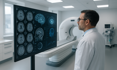
Beyond X-rays, AI extends into the harder studies. On MRI, it supports analysis of soft tissue, cartilage, and the degenerative changes behind chronic joint pain. On CT, it helps map complex and comminuted fractures and build the 3D picture surgeons use for pre-operative planning. Used this way, AI radiology software for hospitals supports the full orthopaedic imaging workload.
Integration With Hospital PACS and Workflow
The best solution barely changes your team's habits. Modern systems connect to your existing PACS through DICOM, so scans flow in and results return without a separate login. A typical workflow looks like this.
- The scan is acquired and sent to the PACS as usual
- The AI reviews the image and flags any suspected findings
- The radiologist reviews, confirms, or overrides the flag
- The final report is signed off by the clinician
Meeting CDSCO Rules for AI Diagnostic Imaging
Clinical software in India sits inside a real regulatory framework. AI software that helps diagnose, monitor, or treat a condition is treated as Software as a Medical Device, and it falls under the Central Drugs Standard Control Organisation and the Medical Devices Rules, 2017. Devices are placed into risk classes, and the obligations around licensing, validation, and documentation rise with that risk. AI tools that analyse imaging data to support active diagnosis generally fall under Class C, the moderate-to-high risk category, which is licensed centrally rather than at the state level.
On 21 October 2025, CDSCO issued a Draft Guidance Document on Medical Device Software. Importantly, this guidance explains how the existing Medical Devices Rules apply to software rather than creating new law, and at the time of writing it remains in draft pending final approval. For a hospital, the practical takeaway is simple. CDSCO compliant AI medical imaging software is software built and documented with that risk-based framework in mind from the start, not bolted on later.
Two points matter for an Indian deployment. First, the radiologist remains the accountable decision-maker, and the AI assists rather than replaces clinical judgment. Second, patient imaging is personal data, governed in India by the Digital Personal Data Protection Act, which carries consent and security obligations. A serious solution respects both. You can review the current framework directly through the regulator at the CDSCO website.
No credible partner can guarantee a regulatory outcome, so be wary of anyone who claims to. Good engineering builds the software the right way so that compliance is achievable.
What Orthopaedic Hospitals Gain From Compliant AI Imaging
The payoff is felt across the hospital. For the medical director, it means fewer missed diagnoses and more consistent reads. For the radiology head, a lighter load and faster turnaround on a heavy day. For the owner or managing director, lower medico-legal risk and a credible claim to diagnostic excellence in a crowded Delhi NCR market.
The gains a speciality hospital can expect include:
- Fewer missed and delayed fracture diagnoses
- Faster reporting and shorter patient turnaround
- Reduced strain on a stretched radiology team
- Lower medico-legal exposure from diagnostic error
- A genuine competitive edge in accuracy and speed
These are not guarantees of perfection. They are realistic improvements that come from giving clinicians a reliable second read. Working with an experienced medical imaging AI development company in India makes those gains far more reachable.
Building a Compliant AI Imaging Solution With the Right Partner
The difference between a promising pilot and a dependable clinical tool is usually the partner behind it. A capable team understands orthopaedic imaging, trains convolutional neural network models through deep learning on relevant, well-labelled data, and integrates the result cleanly into your PACS using the DICOM and HL7 FHIR standards your systems already speak, whether the model runs on cloud or on-premise infrastructure. Crucially, it develops in line with the standards that govern medical device software, IEC 62304 for the software lifecycle and ISO 13485 for quality management. That blend of clinical understanding, recognised quality standards, and engineering discipline is what turns AI medical imaging solutions from a concept into something your radiologists actually trust.
Theta Technolabs works across exactly this space, pairing computer vision expertise with AI-driven digital healthcare solutions built for real clinical settings and compliant deployment in view. If your hospital is exploring how AI could strengthen its imaging, a conversation about your specific workflow is the right place to start, and you can reach the team directly at sales@thetatechnolabs.com.
Frequently Asked Questions
Is AI medical imaging software legal to use in Indian hospitals?
Yes, when it meets the applicable rules. AI imaging software is regulated as Software as a Medical Device under the Medical Devices Rules, 2017, and is placed in a risk class that determines its licensing and validation requirements. A properly developed and compliant solution sits well within the framework.
Does AI replace radiologists for fracture detection?
No. These tools are designed to assist, not replace. The AI provides a second read and flags suspected findings, but the radiologist reviews every result and remains accountable for the final diagnosis.
How accurate is AI at detecting fractures?
Performance varies by the body region, the type of fracture, and the quality of the scan. That is precisely why AI is deployed as a support tool alongside a clinician rather than as a standalone decision-maker. It strengthens the reading process without removing human oversight.
Can it connect to our existing PACS?
In most cases, yes. Modern systems integrate with hospital PACS through the DICOM standard, so the AI fits into your current workflow rather than forcing a separate one.
How is patient imaging data protected?
In India, patient imaging is personal data governed by the Digital Personal Data Protection Act, which sets obligations around consent and data security. A responsible solution is built to honour those requirements.
Conclusion
Diagnostic errors in orthopaedics are costly, but they are not inevitable. With well-built, compliant AI medical imaging solutions, a speciality hospital can give its clinicians a reliable second read, ease the pressure on its radiologists, and protect both its patients and its reputation. The technology is ready, and the right partner makes it work.






.png)





.png)






















.avif)
.avif)
.avif)













_How%20IoT%20Can%20Reduce%20Energy%20Costs%20in%20Smart%20Factories_Q4_25.avif)
_How%20AI%20Development%20Companies%20in%20Ahmedabad%20are%20Transforming%20the%20Shopping%20Experience_Q4_25.avif)
_Node.js%20and%20Blockchain_%20A%20Perfect%20Pair%20for%20Fintech%20Innovation%20in%20Dubai_Q3_24.avif)
_Choosing%20the%20Right%20Computer%20Vision%20Development%20Partner%20in%20Ahmedabad%20for%20Construction_Q3_24.avif)
_The%20Transformative%20Role%20of%20Open%20Banking%20APIs%20in%20Fintech%20for%202024_Q3_24.avif)
_Explore%20the%20Best%20Cross-Platform%20App%20Development%20Frameworks%20of%202024_Q3_24.avif)
_Integrating%20IoT%20with%20Mobile%20Apps%20for%20Advanced%20Renewable%20Energy%20Solutions_Q2_24.avif)
_Top%20Benefits%20of%20Cloud%20Computing%20for%20All%20Business%20Sectors_Q2_24.avif)